News Story
New robust and scalable computational methodology developed by UMD researchers helps identify directed connectivity within the brain
Exciting work from a cross-laboratory collaboration at the University of Maryland has produced a cutting-edge method for directional connectivity with magnetoencephalography (MEG). Dubbed “NLGC”—for Network Localized Granger Causality—the method obtains directional cortical connectivity measures from MEG recordings.
A paper describing the new inference method appears in the October 2022 open-access Elsevier journal Neuroimage.
The research that produced NLGC: Network Localized Granger Causality with Application to MEG Directional Functional Connectivity Analysis was led by first author Behrad Soleimani, a Ph.D. student of Associate Professor Behtash Babadi (ECE/ISR); Professor Jonathan Simon (ECE/ISR/Biology); alumnus Proloy Das (ECE Ph.D. 2020), a former student of Babadi who is currently a research fellow in anesthesia at Massachusetts General Hospital; I. M. Dushyanthi Karunathilake, a Ph.D. student of Simon; Stefanie E. Kuchinsky, a research investigator in the Audiology and Speech Pathology Center, Walter Reed National Military Medical Center and an affiliate of UMD’s Hearing and Speech Sciences (HESP).
Identifying the directed connectivity that underlies networked activity between different cortical areas in the brain is critical for understanding the neural mechanisms behind sensory processing.
“To understand how the brain works, we need to know more than what any one part of the brain is doing,” says Jonathan Simon. “We also need to know how the different parts of the brain give and get information from each other. Many brain areas provide critical support for other brain areas.”
Granger causality (GC) is a data-driven methodology for statistical assessment of directed connectivity that can quantify the flow of information based on improvement in the temporal predictability of a time-series, given the history of another one. GC is widely used for this purpose in functional magnetic resonance imaging (fMRI) analysis, but there the temporal resolution is low, making it difficult to capture millisecond-scale interactions that underly sensory processing. In contrast, MEG has millisecond resolution, but only provides low-dimensional sensor-level linear mixtures of neural sources. This makes using GC inference a challenging task.
Conventionally, methods proceed in two stages. First, cortical sources are estimated from MEG using a source localization technique. This is followed with GC inference among the estimated sources. However, the spatiotemporal biases in estimating sources that propagate into the subsequent GC analysis stage may result in both false alarms and in missing true GC links.
The authors’ new NLGC inference paradigm localizes directional connectivity directly, instead of first localizing sources and then estimating their connectivity. It models the source dynamics as latent sparse multivariate autoregressive processes and estimates their parameters directly from MEG measurements, integrated with source localization, and employs the resulting parameter estimates to produce a precise statistical characterization of detected GC links.
Here, GC is used as a directed connectivity measure, measuring the influence of one cortical source on another if its removal from a predictive model results in worse prediction. The connection can be in either direction, or both directions.
“NLGC addresses the key challenge of computational scalability in connectivity analysis, which is that estimating all possible connections between hundreds to thousands of sources in the brain using previous algorithms is computationally burdensome,” says Behtash Babadi. “By integrating several existing approaches from signal processing and machine learning into a unified framework, NLGC provides several orders of magnitude of saving in computation time.”
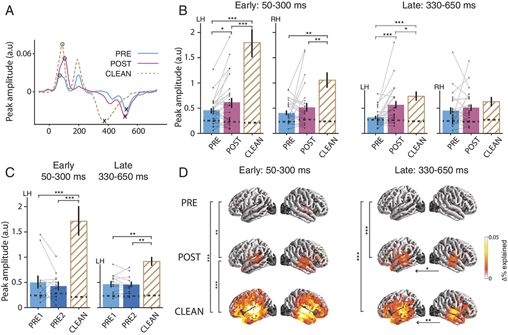
NLGC is able to outperform other methods by avoiding the bias propagation of the two-stage procedure. It is more accurate and able to limit false positives without overly suppressing true positives. NLGC sidesteps typical “current spread” artifacts, provides the direction of the connectivity (not just the strength of the connection), and takes full advantage of the exquisite (millisecond) temporal resolution of MEG. NLGC is markedly robust with respect to model mismatch, network size, and low signal-to-noise ratio; conventional two-stage methods result in high false alarms and misdetections.
“We carefully examined the performance of NLGC in various realistic scenarios using comprehensive simulation studies as well as application to MEG data,” Babadi says. “Due to its usage of a flexible, yet biologically informed statistical model as well as a rigorous statistical testing framework, NLGC stood out among several existing widely-used methods in terms of its accuracy and robustness.”
How NLGC could be used
Researchers will be able to investigate different kinds of brain connectivity in various frequency bands, particularly in auditory tasks, by using NLGC with MEG.
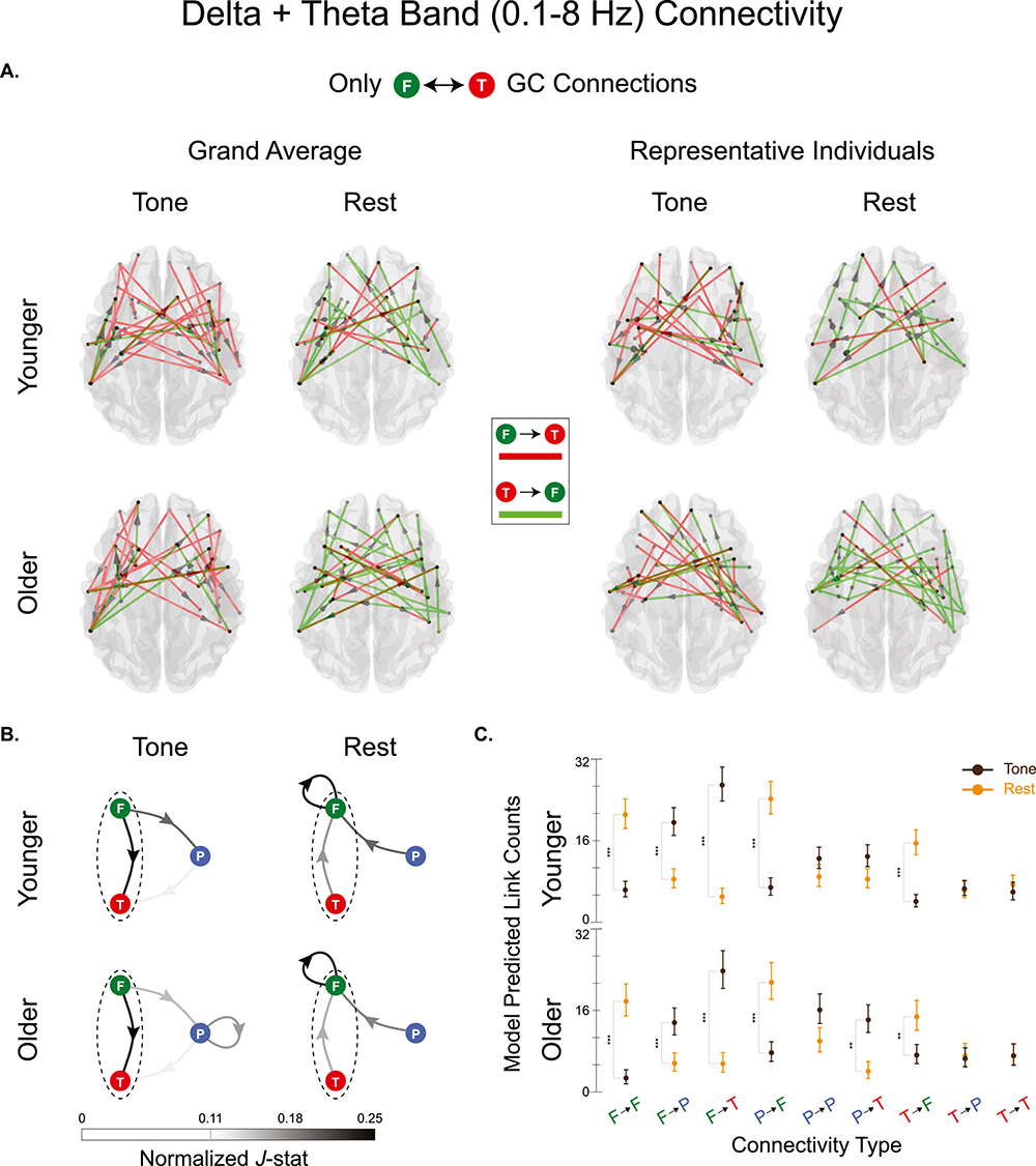
Already, the method has been used to compare which brain area connections are used in an active listening task, compared to when the brain is in a contemplative state (sometimes called the “resting” state, but the brain is never truly at rest). NLGC shows that during active listening, one type of connectivity (“delta band”) is dominantly outward from the Frontal lobe (the area responsible for enforcing the performing of the active task), while the connectivity in the contemplative state has dominated by the Frontal lobe receiving input from the other areas, including many connections from itself.
NLGC has proven so useful it is already being employed in other investigations.
For example, NLGC is helping the researchers show how the brain’s connections differ between older and younger adults. “We are using it in a study of how the Frontal and Parietal lobes help the auditory cortex when a person is listening to people talking in a noisy room,” Simon explains. NLGC is also being used to investigate connectivity changes underlying recovery from stroke. “It’s helping us understand how a stroke can impact the entire brain—not just in the part of the brain where the stroke occurs,” Simon notes. “We are learning how strokes affect how key areas of the brain communicate with each other.”
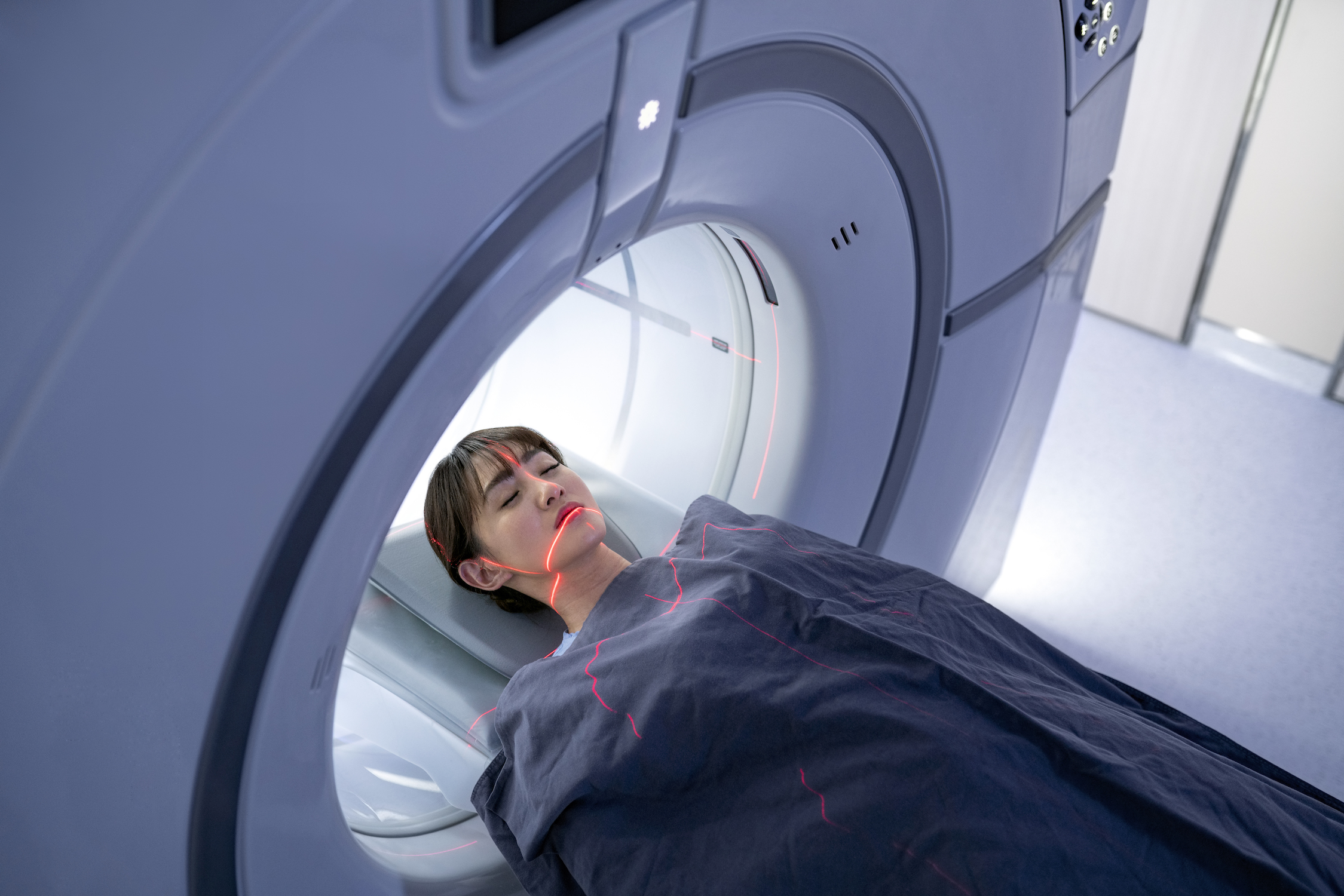
Photo credit for this story's "thumbnail" is Bethany Swain, University of Maryland. Thank you Bethany!
Published September 2, 2022
Related Stories
Stories / September 9, 2022
$7.9 Million in NIH Awards Propel UMD Aging Research
Stories / June 16, 2022
Jonathan Simon gives keynote address at international cognitive...
Stories / May 9, 2022
Poster Session Cinches Banner Year for UMD Neuroscience
Stories / November 1, 2021
How Home Alone Helped UMD Neuroscientists Unlock Brain Scan Data
Stories / July 8, 2021
NIH grant furthers poststroke recovery research of Marsh, Simon

Stories / December 14, 2020
Emerging from the fog: Little understood post-stroke cognitive...

Stories / July 24, 2019
Simon invited speaker at implantable auditory prostheses...
Stories / December 7, 2023
‘Priming’ helps the brain understand language even with...
Stories / September 26, 2023
New UMD Division of Research video highlights work of Simon,...

Stories / August 23, 2023
UMD Researchers to Untangle Language Problems for Tongue-Tied...