News Story
Announcing the BBI Small Animal MR Facility
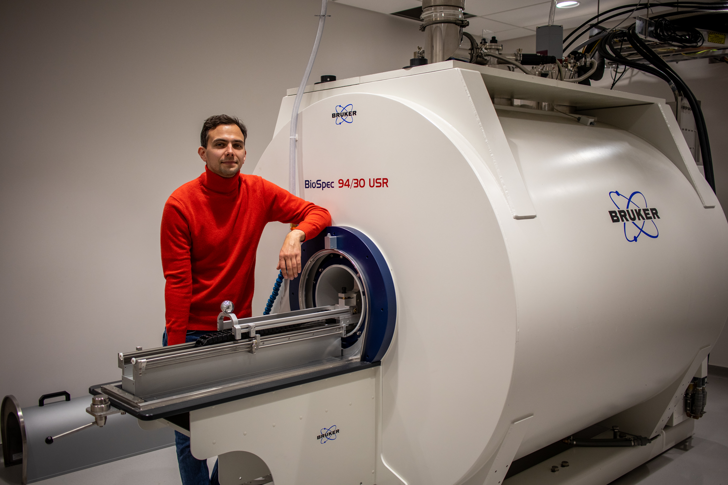
BBI-SAMRI Director Konstantin Cherkas with Bruker BioSpec 94/30 in A. James Clark Hall.
Magnetic resonance (MR) imaging, a cornerstone of non-invasive brain imaging, is now available at the new Brain and Behavior Institute small animal MR facility (BBI-SAMRI). Led by director Konstantin Cherkas, the BBI-SAMRI will expand campus research to allow anatomical, functional and connectomic analysis of the nervous, cardiovascular and musculoskeletal systems.
With a liquid helium-cooled coil (CryoProbe) to boost signal as much as 25-fold and the capacity to image across a range of animal models, the BBI-SAMRI will positively impact research on biological systems across the campus community. Applications include imaging central nervous system development and aging, neuroinflammation, photodynamic therapies and immune engineering.
“We are excited about harnessing the power of the new BBI small animal MR facility and the expertise of director Cherkas to offer a range of new tools to study the structure and function of the nervous system in health and disease,” said Elizabeth Quinlan, Professor of Biology, Clark Leadership Chair in Neuroscience and Director of the BBI. “This facility, like other new BBI research cores, is geared toward tackling grand challenges in neuroscience by expanding research capabilities at UMD.”
The BBI-SAMRI employs the highly versatile Bruker BioSpec 94/30, which boasts 9.4T of imaging power and a 30cm inner gradient diameter with an innovative modular concept for gradient inserts. Located on the first floor of A. James Clark Hall, the facility is ideally situated to promote complementation with imaging modalities on the sixth-floor vivarium, including PET, ultrasound and two-photon microscopy.
The BBI-SAMRI grand opening will take place on Tuesday, April 26, 2022 and feature keynote speaker Dr. Alberto Vazquez from the University of Pittsburgh.
MR imaging can be used to quantify changes in the brain and spinal cord induced over early development, following learning and injury, or with age and disease progression. Although small animal imaging can be performed on MR scanners designed for humans, dedicated small animal systems have significant benefits, particularly superior spatial and temporal resolution. In addition, the preclinical MR imaging offered by the BBI facility will enable the acquisition of longitudinal data, the minimization of experimental subjects, a reduction in biological variability and the promotion of clinical translation.
Cherkas’s vision for the BBI-SAMRI is to make the new facility as user-friendly as possible in terms of scanning experience and post-processing data interpretation. While aspiring to reduce the amount of time spent acquitting the desired images, particularly images of evoked activity, Cherkas also seeks to foster proficiency with post-processing procedures, which can be especially intimidating to new users.
“My overall aim for the BBI-SAMRI is a minimum-click outcome, meaning that researchers can access information in just two or three clicks,” Cherkas said, “Investigators at the facility should be able to acquire and interpret their findings with relative ease. To enable this, I will offer training sessions to introduce users to the theory and practice of small animal MR imaging, consultations on experimental protocols and hands-on training for image acquisition and analysis.“
Cherkas noted that the facility has plenty of appeal for physical scientists as well, as the BBI-SAMRI represents the intersection of biological problems and quantitative methods.
“MR imaging is a field that welcomes mathematicians, physicists, computer scientists and engineers in addition to researchers working in the translational pipeline,” he said. “The BBI-SAMRI represents an opportunity for investigators interested in everything from pulse sequence and hardware development to optimization of data reconstruction algorithms and applications of machine learning across the facility.”
Cherkas earned his bachelor and master of science degrees in nuclear physics from Taras Shevchenko National University in Kyiv, Ukraine, in 2011 and 2012 respectively. In 2017, he received his Ph.D. from the Institute for Nuclear Research in Kyiv, where his dissertation focused on the theoretical and experimental aspects of nuclear processes at the interaction of exotic ions. Cherkas then joined the Center for Neuroscience Imaging Research at the Institute for Basic Science in Suwon, South Korea, where he worked on 9.4T and 15.2T Bruker BioSpec systems. His continued research interests include the development of the MR compatible optical devices, BIDS compatible pipelines for MR data post-processing and machine learning for signal processing.
“I am keen to augment and expand the research of Maryland investigators,” Cherkas noted, “Come to the BBI-SAMRI, and I will help facilitate the proper fit for your work to the optimal imaging modality.”
###
Writer: Nathaniel Underland, underlan@umd.edu
About the Brain and Behavior Institute: The mission of the BBI is to maximize existing strengths in neuroscience research, education and training at the University of Maryland and to elevate campus neuroscience through innovative, multidisciplinary approaches that expand our research portfolio, develop novel tools and approaches and advance the translation of basic science. A centralized community of neuroscientists, engineers, computer scientists, mathematicians, physical scientists, cognitive scientists and humanities scholars, the BBI looks to solve some of the most pressing problems related to nervous system function and disease.
Published January 31, 2022
Related Stories
Stories / February 28, 2022
An Open House Debut for New Prisma MRI at MNC

Stories / February 14, 2022
UMD Chemist Pioneers Tool for Eavesdropping on Neurons
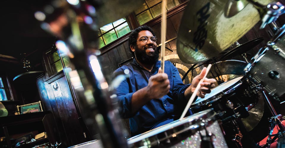
Stories / February 2, 2022
Researcher Beats His Own Drum in Considering How Brains Process...
Stories / December 15, 2021
Neuroscience Graduate Students at UMB, UMCP Awarded Fellowships...
Stories / November 20, 2019
New Perspectives on Some of the Oldest Questions about Human...
Stories / September 30, 2019
NIH announces HEAL Initiative Grants for UMD faculty
Stories / April 28, 2023
Autism Research Resonates in Hearing-Focused Project

Stories / March 6, 2023
Isolated Cells, Inclusive Science
Stories / January 23, 2023
2023 BBI Seed Grants Inspire New Interdisciplinary...
Stories / October 26, 2022
Training Can Improve Older Adults’ Ability to Discriminate...